


Dr Ngaserin Sabrina Ng Hui Na
General Surgeon


 Add as a preferred source on Google
General Surgeon
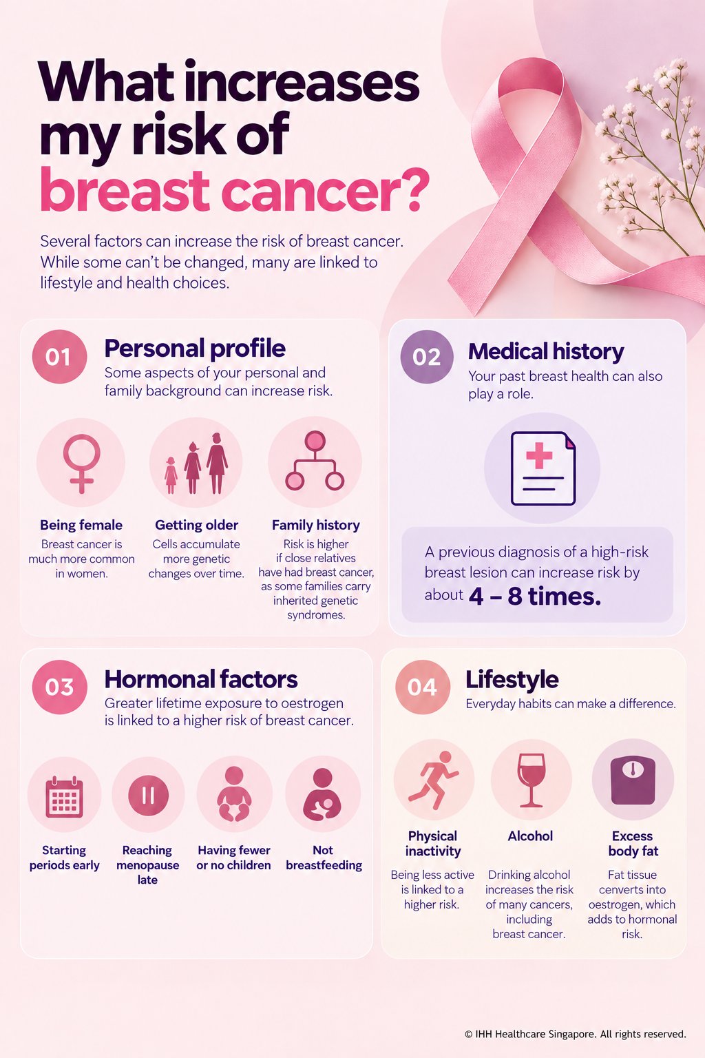
Breast cancer risk is influenced by a combination of non-modifiable, hormonal, genetic and lifestyle factors. Being female and getting older are the two biggest underlying risk factors. Other factors include a family history of breast cancer, inherited gene mutations such as BRCA1 and BRCA2, previous high-risk breast lesions, and greater lifetime exposure to oestrogen, for example, through earlier menstruation or later menopause.
Lifestyle factors can also play a role. Physical inactivity, alcohol consumption and excess body weight, particularly after menopause, are associated with an increased risk of breast cancer.
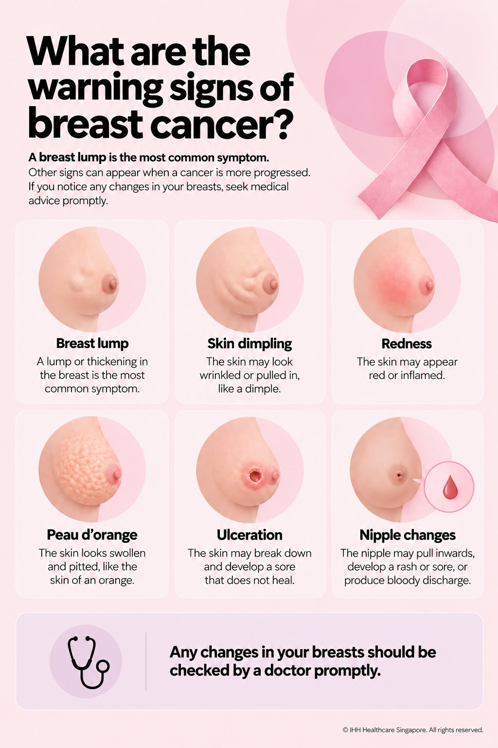
The most common sign of breast cancer is a new breast lump, but breast cancer does not always present with a lump. Other warning signs can include a change in breast shape or size, skin dimpling or thickening, nipple inversion or a new, persistent nipple discharge. Some early breast cancers have no symptoms at all and are detected only through screening.
Breast cancer is the most common cancer in Singapore and around the world - 1 in 12 women in Singapore risk developing the disease by 75 years of age.
Dr Sabrina Ngaserin, a breast surgeon at Mount Elizabeth Hospitals, says the scale of the disease is significant. “Breast cancer accounts for a remarkably large proportion of the cancer burden among women in Singapore. It is not a rare disease – conversely it is something that many families will encounter, directly or indirectly.”
The encouraging news is that more women are being diagnosed earlier, and survival has improved considerably. The age-standardised relative survival rate has risen from 49.9% between 1973 and 1977 to over 83% between 2018 and 2022 (National Registry of Diseases Office 2024). The percentage of breast cancers diagnosed at Stage I in Singapore has increased dramatically from 33% between 2008 and 2012 to over 57% between 2018 and 2022 (National Registry of Diseases Office 2024).
This progress reflects advances across the entire spectrum of breast cancer care -from earlier detection and more precise diagnosis to increasingly effective systemic therapies, surgery and radiotherapy.
“As a doctor, my goal is not simply to treat the cancer, but to give every patient the best possible chance of a long and meaningful life - whether that means curing the disease, controlling it for as long as possible, or helping someone live well through treatment.” says Dr Ngaserin.
“For patients diagnosed with Stage 0 to Stage 3 breast cancer, treatment is generally delivered with curative intent. When cancer has spread to other organs at Stage 4, the goals of treatment change. We focus on containing the disease, managing symptoms, extending meaningful survival and, importantly, helping patients live as well and as fully as possible.”
Yes. Breast cancer can affect women at any age, including during their 20s, 30s and 40s, and it can occur before or after menopause, regardless of breast size or family history. Men can develop breast cancer too, although male cases account for less than 1% of all breast cancers.
Dr Ngaserin notes that she sees breast cancer in younger women, a group in whom symptoms can sometimes be overlooked or attributed to more common benign causes.
She recalls a patient who had a breast lump for more than a year. She first noticed it while breastfeeding her first child, but it was only diagnosed as breast cancer when she became pregnant with her second child.
“One of the challenges with young women is that a breast lump can easily be attributed to breastfeeding, hormonal changes or a benign condition,” says Dr Ngaserin. “But persistence matters. If a lump does not resolve, it deserves proper assessment - regardless of a woman’s age or whether she is breastfeeding.”
The patient was able to receive chemotherapy during pregnancy, followed by breast-conserving surgery after delivering her baby and subsequently radiotherapy. More than 5 years later, she remains well.
Her story highlights an important message: breast cancer may be less common in young women, but it is not impossible. A new or persistent breast change should never be dismissed purely because of age, pregnancy or breastfeeding.
“Breast cancer is not one disease, and treatment is no longer one-size-fits-all. Today, we have more ways to tailor treatment to the biology of the cancer and to the woman herself. The goal is not simply to treat the cancer, but to give each patient the best possible chance of living a long and meaningful life.”
Dr Ngaserin groups breast cancer risk factors into 4 broad areas:
Personal profile: Being female and getting older are the 2 strongest underlying risk factors. As we age, our cells have had more opportunities to accumulate genetic changes. Family history is also important, particularly when a mother, sister or daughter has had breast cancer, especially at a young age or when several close relatives are affected. In some families, this may reflect an inherited genetic predisposition such as a BRCA1 or BRCA2 mutation.
Medical history: Certain previous breast conditions can increase future breast cancer risk. High-risk lesions such as atypical hyperplasia and lobular carcinoma in situ (LCIS) are associated with a substantially increased risk, depending on the specific diagnosis, risk may be several times that of the general population.
Hormonal and reproductive factors: Breast tissue is influenced by hormones, particularly oestrogen and progesterone. A longer lifetime exposure to these hormones, for example, through earlier onset of menstruation or later menopause, is associated with increased breast cancer risk. Conversely, pregnancy and breastfeeding are associated with a modest reduction in risk. These are, of course, deeply personal life choices and should never be viewed simply as breast cancer prevention strategies.
Lifestyle and metabolic factors: Physical inactivity, alcohol consumption and excess body weight, particularly after menopause, are associated with increased breast cancer risk. After menopause, fat tissue becomes an important source of oestrogen, which is one reason excess body fat is linked to increased risk of hormone-sensitive breast cancers. Maintaining a healthy weight and staying physically active can therefore contribute to overall breast health as well as general wellbeing.
“Risk is rarely about one factor alone,” says Dr Ngaserin. “It is usually a combination of our genetics, biology, medical history and environment. The important thing is to understand your individual risk, because that allows us to decide whether you need a different or earlier screening strategy.”
Validated breast cancer risk calculators can estimate a woman’s 5-year and lifetime risk of developing breast cancer, based on factors such as age, family history, reproductive history, previous breast conditions and, in some models, breast density and genetic information. Different calculators use different factors and may therefore produce different estimates for the same woman.
Dr Ngaserin uses internationally validated risk assessment tools as a guide. “As a general guide, a lifetime risk below 15% is usually considered average risk, while a lifetime risk of 20% or more is generally considered high risk and may warrant specialist assessment and enhanced screening, depending on the individual’s risk factors.”
The American Cancer Society recommends annual breast MRI in addition to mammography for women with a lifetime risk of approximately 20 to 25% or greater, although the exact approach should be individualised.
“Many women fall into an intermediate range between these figures,” says Dr Ngaserin. “This is often where things become less straightforward. A risk estimate is not a diagnosis, and it does not tell us exactly what will happen to an individual. For women in this intermediate-risk group, particularly those with a significant family history or previous high-risk breast lesions, a breast specialist can put the number into context and advise on the most appropriate screening strategy.”
People with a significantly elevated risk of breast cancer, particularly when there is a strong family history or suspicion of an inherited cancer syndrome, may be offered genetic counselling and testing.
At Mount Elizabeth Hospitals, genetic assessment can involve a multidisciplinary team, which may include genetic counsellors, medical oncologists and other relevant specialists. Counselling before testing helps determine whether genetic testing is appropriate and which test is most informative. After testing, the team explains the results and what they may mean for the patient and, where relevant, their family members.
Genetic test results are generally reported in 3 broad categories:
Positive: A pathogenic or likely pathogenic variant associated with an increased risk of cancer has been identified. The specific gene involved helps determine what the result means for the individual and whether additional screening, risk-reducing strategies or testing for family members should be considered.
Negative: No pathogenic or likely pathogenic variant was identified in the genes examined. This can be reassuring, but it does not necessarily mean that a person has no increased risk. If there is a strong family history or other risk factors, the person's overall risk may still be elevated even when genetic testing is negative.
Variant of uncertain significance (VUS): A genetic change has been identified, but there is currently insufficient evidence to determine whether it increases cancer risk. A VUS is not considered a positive result and should generally not be used on its own to guide major medical decisions. As scientific knowledge evolves, the classification may be updated.
The main inherited genetic syndrome associated with breast cancer is hereditary breast and ovarian cancer (HBOC) syndrome, most commonly caused by pathogenic variants in the BRCA1 or BRCA2 genes.
“These mutations are found in only a minority of people with breast cancer,” explains Dr Ngaserin. “Around 5–10% of breast cancers are associated with inherited pathogenic variants in BRCA1 or BRCA2. The majority of breast cancers are not caused by an inherited mutation; they arise through genetic changes acquired during a person’s lifetime, influenced by a combination of ageing, biology and environmental factors.”
For women who inherit a pathogenic BRCA1 or BRCA2 variant, however, the risk is substantially higher than in the general population. Current estimates suggest a lifetime breast cancer risk of approximately 47–66% for BRCA1 and 40–57% for BRCA2, although estimates vary between studies and populations.
BRCA1 and BRCA2 variants can also increase the risk of other cancers, including ovarian, fallopian tube, primary peritoneal, melanoma, pancreatic and prostate cancers, as well as male breast cancer. The strength of the association varies by gene and cancer type.
For people found to carry a pathogenic BRCA1 or BRCA2 variant, management is individualised. Options may include more intensive breast surveillance, medication to reduce breast cancer risk in selected women, and risk-reducing surgery such as bilateral mastectomy or removal of the ovaries and fallopian tubes. These are highly personal decisions and depend on age, family plans, previous cancer history, the specific gene involved and the individual's preferences and risk tolerance.
“The value of knowing your genetic risk is not simply knowing a number,” says Dr Ngaserin. “It gives us an opportunity to be proactive – for us to tailor screening, consider risk-reducing options where appropriate, and potentially identify risks that are relevant not only to the patient, but also to her family.”
A new breast lump is the most common symptom of breast cancer, but it is not the only warning sign.
When a cancer develops close to the nipple, the nipple may pull inwards (become inverted) or produce spontaneous, persistent or bloody discharge. The nipple or surrounding skin may also develop a rash, sore or persistent change in appearance.
As breast cancer progresses, other changes may appear, including skin dimpling, redness, thickening or ulceration. Another recognised sign is peau d’orange, where the skin becomes swollen and pitted, taking on the appearance of an orange peel.
Importantly, breast cancer does not always cause obvious symptoms. Some cancers are found through screening before a woman can feel or see any change.
The message is simple: know what is normal for your breasts, and don’t ignore a change that is new or persistent. If something feels or looks different, have it checked promptly.
A breast specialist will usually begin with a thorough clinical examination and an assessment of your individual risk factors. This is followed by appropriate breast imaging, which may include a mammogram, ultrasound and, in selected situations, MRI. If an abnormality is identified, a tissue biopsy may be recommended to determine exactly what it is.
If cancer is confirmed, the biopsy tells your doctor several important things:
The type of cancer: The most common invasive breast cancer is invasive ductal carcinoma, which begins in the milk ducts, followed by invasive lobular carcinoma, which starts in the milk-producing lobules.
The grade: Breast cancers are generally graded from 1 to 3, based on how abnormal the cancer cells look under the microscope and how closely they resemble normal breast cells. A higher grade generally indicates a cancer with more aggressive biological features.
The receptor status: The cancer cells are tested for oestrogen receptors (ER), progesterone receptors (PR) and HER2. This helps us understand the biology of the cancer and, importantly, guides treatment. Hormone receptor-positive breast cancers are the most common and often can often be treated effectively with the inclusion of hormone therapy. HER2-positive and triple-negative breast cancers have different biological characteristics and may benefit more greatly from specific systemic treatments, including chemotherapy, targeted therapy or immunotherapy in appropriate patients.
Once a diagnosis has been established, further imaging may be recommended depending on the type and stage of cancer. MRI, CT, FDG-PET/CT or bone imaging may be used in selected patients to assess the extent of disease and determine whether there is evidence of spread beyond the breast and regional lymph nodes.
A biopsy does much more than confirm that a cancer is present - it gives us the biological information we need to understand the cancer and tailor treatment to the individual patient.
Often, yes.
The cells lining the breast ducts naturally renew themselves throughout our lives. Sometimes, these cells begin to multiply more than usual, a process known as hyperplasia, which is still benign. If additional mutational changes occur, the cells may develop abnormal features, known as atypical hyperplasia. These cells are not cancer, but they represent a higher-risk change and an opportunity for early intervention.
In some women, further changes may lead to ductal carcinoma in situ (DCIS) i.e. Stage 0 breast cancer, where abnormal cells remain confined within the breast duct and have not yet invaded the surrounding tissue. If these abnormal cells eventually breach the duct wall, they can develop into invasive breast cancer, which can range from Stage 1 through Stage 4 depending on how far the disease has spread.
“Breast cancer does not develop overnight. it is a process, and there are usually opportunities to intervene along the way,” says Dr Ngaserin. “The earlier we identify high-risk changes or cancer, the more options we may have to intervene effectively. In some situations, early intervention of pre-cancerous changes may even prevent breast cancer from developing in the first place.”
Treatment is carefully planned by a multidisciplinary team (MDT) of specialists, which may include breast surgeons, medical and radiation oncologists, specialist nurses, physiotherapists, psychologists and genetic counsellors. Bringing different perspectives together allows the team to tailor treatment not only to the cancer, but also to the individual patient.
The sequence of treatment matters. For many patients with early-stage breast cancer, surgery is the first step, followed by treatments such as radiotherapy, chemotherapy, targeted therapy or hormone therapy where appropriate.
However, this is not always the case. Depending on the size and biology of the tumour, lymph node involvement and other individual factors, some patients may receive systemic treatment, such as chemotherapy, targeted therapy or immunotherapy – before surgery. This is known as neoadjuvant treatment and can shrink the tumour, treat microscopic disease early and, in selected patients, make breast-conserving surgery possible or improve the chances of achieving a complete response.
Equally important is what matters to the patient.
“Breast cancer treatment is not a one-size-fits-all pathway,” says Dr Ngaserin. "The most important thing in my book is understanding the woman behind her diagnosis – her personal goals, priorities and I always respect the element of choice.”
“A woman who is single, newly married, building her career or planning a family may have very different concerns from someone who is retired. Fertility, body image, sexual health, menopause, work, family responsibilities and quality of life can all influence how we approach treatment.”
Not necessarily.
The 2 main types of breast cancer surgery are mastectomy, which removes the whole breast, and breast-conserving surgery, which removes only part of it. Most patients diagnosed early who wish to keep their healthy breasts are eligible for breast-conserving surgery.
Safety in removing the cancer always comes first. The first priority is to achieve sound cancer treatment and, where appropriate, a clear margin of removal. Once oncological safety is established, we can consider the range of surgical options and what is right for the individual patient. To plan surgery, your doctor will consider the size of the tumour relative to the breast volume, where it sits, and the patient's breast shape and overall physical form.
“For example, a tumour taking up around 10% of the breast can usually be removed easily,” Dr Ngaserin says. “A tumour taking up 20–30% or even 50% may require oncoplastic breast surgery, breast remodelling, reduction or partial breast reconstruction.”
Some women may choose a mastectomy without reconstruction. For some, the appeal is simplicity, avoiding additional reconstructive surgery and the need for implants or tissue transfer. A well-fitted external breast prosthesis can also provide a natural shape under clothing.
However, removing one breast can alter the body's symmetry and weight distribution. For some women, this may affect posture, shoulder mechanics or body balance over time, although the impact varies from person to person. This is one of the factors worth discussing when considering the long-term implications of different surgical options, not simply how the breast will look immediately after the operation.
Oncoplastic breast surgery combines cancer surgery with plastic surgical techniques to remove the tumour safely while preserving, or restoring, the natural shape and appearance of the breast. The aim is to achieve good oncological outcomes without unnecessarily compromising how the breast looks and feels afterwards.
Minimally invasive breast surgery takes this principle further by using endoscopic or robotic techniques through smaller, strategically placed incisions to perform nipple and skin sparing mastectomy. Depending on the procedure and the individual patient, small 2-4 cm scars may be positioned in less conspicuous areas such as the armpit or along the bra line. The same incisions can be used to perform whole breast reconstruction.
“Breasts are a very private part of the body, but they are also something women see every day - in the mirror, when they shower and when they get dressed,” says Dr Ngaserin. “A scar or change in breast shape may seem like a small thing from a purely surgical perspective, but for the woman living with it, it can be a daily reminder of what she has been through. Whenever it is oncologically safe to do so, I believe we should think beyond simply removing the cancer and consider how we can help a woman feel comfortable and confident in her body afterwards.”
After a mastectomy, the breast can be reconstructed in several ways, depending on a woman’s anatomy, previous treatments, lifestyle and personal preferences. Options include:
Implant-based reconstruction – using breast implants to recreate the breast shape, with or without acellular dermal matrix.
Latissimus dorsi reconstruction – using muscle and tissue from the back, sometimes combined with an implant. This can be done with a minimally invasive single-incision, single-port approach with an incision in the underarm.
Abdominal tissue reconstruction – using a woman’s own tissue from the abdomen to create a new breast, frequently performed as a free perforator flap, such as a DIEP flap.
Fat grafting – using the patient’s own fat to restore volume, refine contours or improve symmetry, often as an additional step rather than a complete reconstruction on its own.
Other niche techniques include: Omental flap reconstruction, Using the thigh or buttock tissue for reconstruction, and more.
Reconstruction is also evolving beyond simply recreating the breast’s shape. Patients may also choose to perform sensory preserving mastectomies with nerve reconstruction or neurotisation - to reconnect or preserve sensory nerves, with the aim of improving the return of breast or nipple sensation.
“The goal of reconstruction is not simply to replace what has been removed,” says Dr Ngaserin. “We are increasingly thinking about the breast in multi-dimensions - its shape, contour, softness and, where possible, its sensation. Reconstruction should be personalised to what matters most to each woman.”
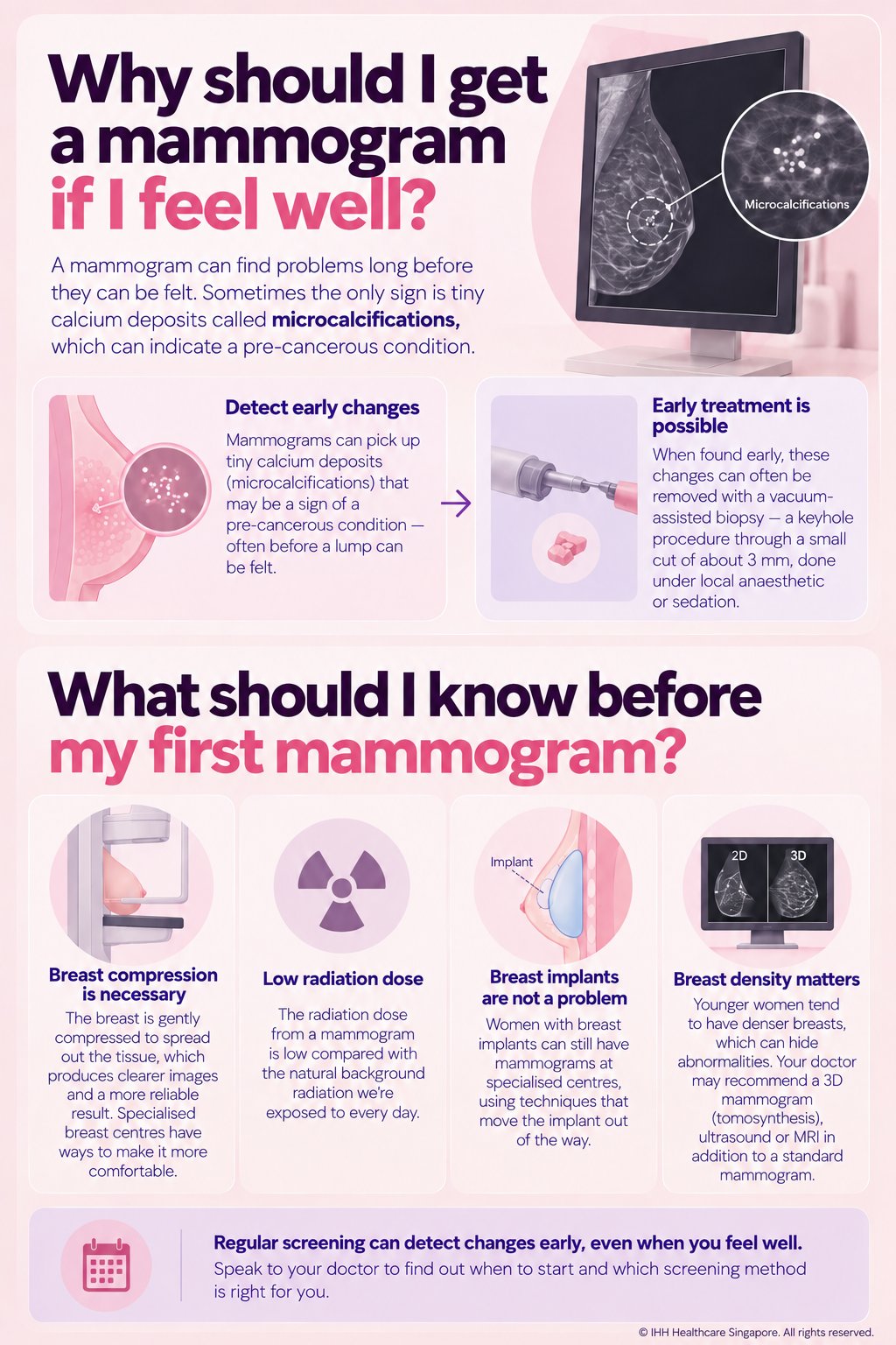
A mammogram can detect breast abnormalities long before they can be felt. Sometimes, the only indication of an early abnormality is a cluster of tiny calcium deposits known as microcalcifications on mamamogram or indeterminate breast lesions later confirmed on ultrasound. While many microcalcifications are benign, certain patterns can be associated with Atypical Hyperplasia or early stage breast cancer as ductal carcinoma in situ (DCIS), and may require further assessment and management.
When a small or localised abnormality needs to be investigated, vacuum-assisted biopsy (VAB) offers a minimally invasive way to obtain multiple tissue samples through a tiny skin incision, typically around 3 mm, using image guidance and local anaesthesia, with sedation in selected cases.
For carefully selected small lesions, vacuum-assisted excision (VAE) can go a step further, allowing the lesion to be removed almost entirely through the same minimally invasive approach. This can sometimes provide both diagnosis and treatment without the need for a conventional surgical incision.
Compression is an important part of mammography because it spreads the breast tissue into a thinner, more even layer, allowing small abnormalities to be seen more clearly and reducing the chance that they are hidden by overlapping tissue. Although the compression can be uncomfortable, it is brief, and specialised breast centres can use techniques to make the experience more tolerable.
The radiation dose from a modern mammogram is very low. For perspective, the radiation exposure from a standard mammogram is often described as being in the same general range as the additional cosmic radiation received during a long-haul flight, such as a transatlantic round trip. The radiation risk from mammography is therefore very small, and for women undergoing screening, the benefits of detecting breast cancer early generally outweigh this small potential risk.
Women with breast implants can still have mammograms at specialised centres, using techniques that move the implant out of the way i.e. implant-displacement views.
Unregulated or illegal silicone injections into the breast are different. Injected silicone can cause inflammation, scarring and changes in the breast tissue that may make abnormalities significantly more difficult to interpret on imaging. Dr Ngaserin strongly advises against illegal procedures.
Breast density is another important consideration. Younger women are more likely to have dense breast tissue, which can make some abnormalities harder to detect on a standard mammogram. Depending on a woman’s age, breast density, personal risk factors and mammogram findings, her doctor may recommend additional imaging such as 3D mammography (tomosynthesis), ultrasound or, for selected higher-risk women, MRI. These tests are complementary rather than interchangeable, and the appropriate approach should be individualised.
There is no single way to prevent breast cancer, but there are things you can do to reduce your risk and improve the chances of detecting it early.
Maintain a healthy weight, exercise regularly and limit alcohol intake. Having children and breastfeeding are also associated with a lower lifetime risk of breast cancer, although these are personal life choices and are not factors that every woman can or should control.
Know what is normal for your breasts. A formal monthly breast self-examination is useful to help one develop an awareness of how your breasts normally look and feel. Be alert to a new lump, persistent change in shape or texture, skin changes, nipple inversion or spontaneous nipple discharge, particularly bloody discharge, and seek medical attention if you notice something unusual.
Most importantly, understand your personal risk and follow an appropriate screening plan. Women with a strong family history, a known genetic mutation or other risk factors may need earlier or additional screening with specialist guidance.
Dr Ngaserin advises: “Know your risk. Know your breasts. Don’t ignore changes. Live well, screen appropriately, and remember that screening is something we do while we are healthy, not because we are sick.”
Whether you'd like to understand your risk, need a breast lump checked, or are exploring your treatment options, our breast surgeons, oncologists and genetic counsellors work together to guide you, with care shaped around your needs and priorities.
Learn more
A monthly breast self-examination 7 – 10 days after your period starts can save your life.

For those who may be planning on having a baby only after they step over the magic number of 40, here are some issues to consider.

Dr Kelly Loi discusses fibroids, how they affect fertility in women, and the ways to identify, evaluate and manage fibroids.

A breast cancer diagnosis does not mean having to lose your breast, now that many treatment options are available.

Eight myths you might believe about breast cancer screening, and the truth about it.

More women are being claimed by breast cancer now than before. Love them by encouraging them to go for breast screening.

Our breast surgeon explains how to understand your risk, warning signs to know, why screening matters even when you feel well, and what surgery can look like today.

As knee osteoarthritis progresses, everyday activities can become more difficult. Our pain specialist and orthopaedic surgeon explain the treatment options available and how to find the right approach for you.

Dr Edwin Chng from Parkway Shenton explains why weight loss can slow down during medical weight management, and when it may be time to review your treatment plan.
Prostate cancer is common, often silent, and still diagnosed too late in Singapore. Urologist Dr Tan Yung Khan on what every man should know about screening, diagnosis, and treatment.

We answer common questions about haze, and how this can affect common chronic lung conditions such as asthma and chronic obstructive pulmonary disease (COPD), and 8 simple tips to protect ourselves against it.

Our neurosurgeon, Dr Vincent Ng, explains the factors specialists consider when deciding whether a brain aneurysm should be monitored or treated, and what the different treatment options involve.
Gleneagles Hospital is part of IHH Healthcare, one of the world's largest integrated healthcare networks.

Download the MyHealth360 app to access an array of medical services conveniently.


Get trusted medical advice from our specialists, dietitians and physiotherapists directly in your inbox.
Subscribe