Dr Foo Siang Shen Leon
Bác Sĩ Phẫu Thuật Chấn thương Chỉnh Hình


Nguồn: Shutterstock
Bác Sĩ Phẫu Thuật Chấn thương Chỉnh Hình
Vừa mới bị ngã và gãy xương? Có một cảm giác đau nhức khó chịu ở một trong các khớp của bạn? Có một chấn thương liên quan đến thể thao hoặc tình trạng xương không thể biến mất?
Khi bạn đến gặp bác sĩ để được chẩn đoán, họ có thể khuyên bạn thực hiện một trong số vài phương pháp chụp ảnh xương khác nhau. Dưới đây là 3 phương pháp hàng đầu để chẩn đoán vấn đề xương hoặc tình trạng khớp.
Hãy nói chuyện với một bác sĩ phẫu thuật chỉnh hình nếu bạn lo lắng về sức khỏe xương hoặc khớp của mình.
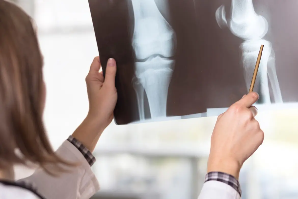
Chụp X-quang sử dụng các sóng năng lượng điện từ, đi thẳng qua cơ thể bạn, để chụp ảnh xương và các cơ quan. Vì mỗi khu vực của cơ thể bạn hấp thụ năng lượng một cách khác nhau, xương của bạn sẽ xuất hiện màu trắng trên hình ảnh X-quang, trong khi các mô mềm sẽ hiển thị các sắc độ của màu xám. Đừng hoảng sợ nếu phổi của bạn trông màu đen – đó là vì chúng chứa không khí!
Chụp X-quang có rất nhiều ứng dụng. Nếu bạn bị thương và bác sĩ nghi ngờ bạn bị gãy xương, bạn sẽ cần một chụp X-quang để xác nhận điều này và xác định mức độ tổn thương.
Bác sĩ của bạn cũng có thể đề nghị chụp X-quang nếu họ nghi ngờ:
Bất kỳ ai cũng có thể chụp X-quang. Nếu bạn đang cảm thấy đau ở một trong các xương hoặc khớp của mình, hoặc bạn bị thương, bác sĩ của bạn có thể khuyên bạn nên chụp X-quang.
May mắn thay, chụp X-quang là thủ tục tiêu chuẩn mang lại rủi ro tối thiểu. Mặc dù chúng sử dụng bức xạ ion hóa, nhưng mức độ phơi nhiễm được coi là an toàn cho hầu hết người lớn. Ngoại lệ duy nhất là nếu bạn đang mang thai, vì bức xạ có thể gây hại cho em bé của bạn. Hãy nói chuyện với bác sĩ của bạn nếu bạn đang mong đợi một đứa trẻ và họ sẽ có thể đề xuất một thủ tục thay thế.
Một tấm ảnh chụp đặc biệt sẽ chụp hình ảnh cơ thể bạn. Bạn sẽ được hỗ trợ vào đúng vị trí, có thể là ngồi, đứng hoặc nằm xuống. Nếu vị trí đó liên quan đến việc di chuyển một xương hoặc khớp bị thương, bạn có thể cảm thấy một số khó chịu trong quá trình kiểm tra. Nếu bạn lo lắng về điều này, bác sĩ của bạn sẽ có thể kê đơn thuốc giảm đau trước.
Bạn cần phải giữ yên trong quá trình chụp X-quang để đảm bảo bác sĩ của bạn nhận được hình ảnh rõ ràng nhất có thể. May mắn thay, việc chụp X-quang thường rất nhanh và hình ảnh thường được chụp trong một phần giây.
Không – chỉ cần nhớ cởi đồ trang sức và bất kỳ vật dụng kim loại nào vì chúng có thể chặn bức xạ X-quang không đi qua cơ thể bạn và làm mờ hình ảnh!
Chụp CT sử dụng sự kết hợp giữa máy chụp X-quang và máy tính tiên tiến để tạo ra một hình ảnh 3D của cơ thể bạn. Chúng cung cấp hình ảnh chi tiết hơn nhiều so với chụp X-quang thông thường, hiển thị cả các cơ quan rắn, mô mềm và bất kỳ sự phát triển bất thường nào cũng như xương của bạn.
Bạn có thể cần chụp CT nếu bác sĩ muốn kiểm tra xương của bạn một cách chi tiết hơn.
Các tình trạng ảnh hưởng đến cột sống, như cong vẹo cột sống (scoliosis) hoặc gãy xương cột sống, có thể cần chụp CT để bác sĩ có thể thấy toàn bộ khu vực bị ảnh hưởng. Bác sĩ của bạn cũng có thể khuyên bạn chụp CT để phát hiện các khối u không ung thư (như u nang) hoặc khối u ung thư, hoặc để đo mật độ xương của bạn (để xác định mức độ nghiêm trọng của loãng xương).
Chụp CT có thể được sử dụng trên hầu như bất kỳ bộ phận nào của cơ thể, từ đầu và vai đến đầu gối và chân, để chẩn đoán các rối loạn xương, gãy xương hoặc ung thư xương. Vì đây là một thủ tục ít xâm lấn, có ít rủi ro ngoại trừ một số phơi nhiễm với bức xạ ion hóa.
Bạn cần nằm xuống để chụp CT. Sau đó, bạn sẽ được vận chuyển vào máy quét, nó sẽ quay xung quanh bạn và chụp hàng trăm bức ảnh, trong vài phút.
Tùy thuộc vào khu vực cơ thể được kiểm tra, bác sĩ của bạn có thể cần sử dụng một loại thuốc nhuộm đặc biệt gọi là chất tương phản để giúp các cơ quan nội tạng, mô và mạch máu của bạn hiển thị rõ nét hơn trong hình ảnh cuối cùng. Bạn thường có thể uống một chất lỏng vô hại chứa chất này. Thay vào đó, bác sĩ đôi khi có thể tiêm nó vào cánh tay của bạn hoặc đưa vào qua đường trực tràng.
Nếu thủ tục của bạn yêu cầu chất tương phản, bạn có thể cần tránh ăn trong khoảng thời gian lên đến 6 giờ trước khi thực hiện thủ tục. Ngoài ra, chụp CT không khác biệt nhiều so với việc chụp X-quang (ở trên) - chỉ là mất thêm một chút thời gian!
Chụp MRI là một thủ tục không xâm lấn kết hợp nam châm lớn, máy tính và sóng từ trường để chụp hình ảnh xương và các cơ quan của bạn. Chụp MRI khác với chụp CT ở chỗ nó mất nhiều thời gian hơn, nhưng lợi ích là hình ảnh có chi tiết phóng xạ học nhiều hơn và quan trọng nhất, nó không phơi bạn dưới bức xạ ion hóa.
Bác sĩ của bạn có thể sử dụng chụp MRI để kiểm tra xương, khớp, sụn, cơ và gân của bạn. Vì vậy, nếu bạn đang gặp phải cơn đau không giải thích được ở bất kỳ khu vực nào trong cơ thể của bạn, bác sĩ có thể khuyên bạn nên thực hiện chụp MRI để xác định nguyên nhân.
Chụp MRI cũng có thể được sử dụng để chẩn đoán:
Vì chụp MRI không sử dụng bức xạ ion hóa, nói chung được coi là an toàn cho hầu hết mọi người, bao gồm phụ nữ mang thai sau tam cá nguyệt thứ nhất, cũng như trẻ em. Tuy nhiên, chụp MRI sử dụng nam châm, vì vậy bệnh nhân có máy tạo nhịp tim hoặc bất kỳ cấy ghép kim loại nào (tùy thuộc vào nhãn hiệu và mẫu của thiết bị) có thể không thể thực hiện thủ tục.
Nếu bạn mắc chứng kín đáo, hãy nói với bác sĩ của bạn về việc dùng thuốc chống lo âu trước khi thực hiện. Đối với bệnh nhân mắc chứng kín đáo nghiêm trọng, cũng có tùy chọn gây mê được giám sát bởi bác sĩ gây mê trong quá trình chụp.
Bạn có thể đã thấy máy MRI trên TV hoặc trong phim – một số người nghĩ chúng trông đáng sợ, nhưng thực sự không cần phải hoảng sợ. Chỉ cần nằm xuống trên giường quét, nó sẽ vận chuyển bạn vào bên trong máy, và hãy giữ cực kỳ yên lặng để máy có thể chụp hình ảnh rõ ràng của bạn. Nhân viên kỹ thuật MRI của bạn sẽ có thể giao tiếp với bạn qua hệ thống liên lạc nội bộ, vì vậy bạn có thể cho họ biết nếu bạn cảm thấy khó chịu hoặc muốn dừng chụp.
Chụp MRI có thể kéo dài từ 20 phút đến 2 giờ, tùy thuộc vào số lượng khu vực cần chụp.
Giống như chụp CT, bạn sẽ không cần phải chuẩn bị nhiều trước khi chụp MRI. Tuy nhiên, bạn sẽ cần phải tháo bỏ mọi trang sức hoặc vật dụng kim loại - đặc biệt quan trọng vì máy sử dụng nam châm. Bác sĩ của bạn cũng có thể phải tiêm thuốc nhuộm tương phản theo cách tương tự như chụp CT.
Không chắc liệu bạn có cần một trong những loại chụp quét được liệt kê ở trên không? Hãy hẹn gặp bác sĩ để tìm hiểu thêm về tình trạng sức khỏe của bạn.